
Article Categories
- All Categories
-
 Data Structure
-
 Networking
-
 RDBMS
RDBMS
-
 Operating System
-
 Java
Java
-
 MS Excel
-
 iOS
iOS
-
 HTML
HTML
-
 CSS
CSS
-
 Android
-
 Python
-
 C Programming
-
 C++
C++
-
 C#
C#
-
 MongoDB
-
 MySQL
MySQL
-
 Javascript
-
 PHP
PHP
-
 Economics & Finance
What Is the Difference Between Organogenesis and Histogenesis
Introduction
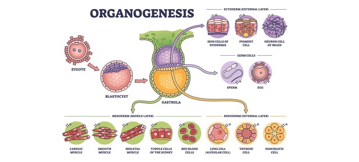
The process of developing organs from three germ layers is called organogenesis. Cell-cell communication, cell fate determination, cell survival and proliferation, cell and tissue size and shape, and the organization of cells into tissues and ultimately functioning organs are all covered.
The process through which cells in an embryo's initial germ layers specialize and take on the properties of the tissues that they eventually give rise to is called histogenesis.
Research in regenerative biology is based on the study of organogenesis since the production of cells and tissues in vivo and in vitro frequently uses regulatory processes that underlie organogenesis.
Cellular and tissue levels can both show signs of histogenesis. Histogenesis occurs at the cellular level, where an early mesoderm cell gradually transforms into a muscle cell.
The majority of the time, however, single cells do not go through histogenesis; instead, they only undergo change when they are a part of a larger cell population that is acting at the tissue level, such as when thousands of cells come together to form the islets of Langerhans tissue in the pancreas.
This tissue is responsible for producing insulin. Organogenesis is the process through which a collection of undifferentiated cells develops into an organ.
Differences Between Organogenesis and Histogenesis
Organogenesis |
Histogenesis |
|---|---|
The stage of embryonic development known as organogenesis begins at the conclusion of gastrulation and lasts until delivery. |
Histogenesis is the process by which undifferentiated cells develop into various tissues. |
The internal organs of the organism are created during organogenesis from the three germ layers that result from gastrulation (the ectoderm, endoderm, and mesoderm). |
These cells are parts of the endoderm, mesoderm, and ectoderm, the three basic germ layers. Histology is the study of the microscopic tissue structures that result from histogenesis. |
The brain and spinal cord are created by the neural tube's growth. Vertebrates have a neural crest that develops into a variety of structures, such as bones, muscles, and central nervous system parts. |
The inner layer of the gastrula, which transforms into the endoderm, is formed by cells that migrate inward along the archenteron. |
The blood, heart, kidney, muscles, and connective tissues of the embryo are formed by the mesoderm, or middle germ layer. |
Fluid cushions and shields the organs from shocks as they develop freely and independently of the body wall inside a coelom. |
Organogenesis
The stage of embryonic development known as organogenesis begins at the conclusion of gastrulation and lasts until delivery. Differentiation, a process whereby less-specialized cells become more-specialized through the expression of a particular set of genes, occurs in each of the three germ layers' cells.
Cell differentiation is driven by cell signaling cascades. Extracellular signals, such as growth factors, which are exchanged with nearby cells in a process known as juxtracrine signaling or with nearby cells within close proximity in a process known as paracrine signaling, affect differentiation.
Intracellular signals, which include cellular signalling (autocrine signalling), are involved in the development of organs.
These signaling pathways enable cell reorganization and guarantee that organs develop at particular locations throughout the organism. Organoids and embryos can be used to study the organogenesis process. The endoderm, which is the embryo's deepest germ layer, develops into the pancreas, liver, and other digestive and respiratory organs by creating their epithelial linings.
The blood, heart, kidney, muscles, and connective tissues of the embryo are formed by the mesoderm or middle germ layer. The developing embryo's ectoderm, or outermost germ layer, gives rise to the epidermis, brain, and nervous system.
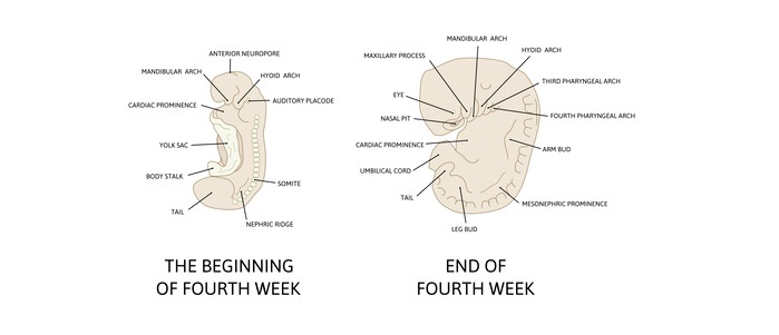
The development of the notochord, which triggers the formation of the neural plate and ultimately the neural tube in vertebrate development, is a key phase in organogenesis for chordates. The brain and spinal cord are created by the neural tube's growth. Vertebrates have a neural crest that develops into a variety of structures, such as bones, muscles, and central nervous system parts.
Often referred to as neurulation, the embryo during this period is known as the neurula and begins to differentiate the ectoderm into the neural crest, neural tube, and surface ectoderm. As the mesoderm splits along the somite axis, the body's coelom develops.
Histogenesis
The process through which cells in an embryo's initial germ layers specialize and take on the properties of the tissues they will eventually give rise to is called histogenesis. A group of cells known as a germ layer are generated during animal and mammalian development.
Within vertebrate creatures, germ layers are often noticeable; nevertheless, animals like mammals that are more complicated than sponges create two or three major tissue layers.
The two layers that radially symmetric animals like cnidarians develop are referred to as the ectoderm and endoderm.
Diploblastic describes them. Animals that have bilateral symmetry create a mesoderm layer as a third layer in between, making them triploblastic. Organogenesis is the process through which germ layers finally give rise to all the tissues and organs of an animal or mammal.
One of the germ layers produced during animal development is the endoderm. The inner layer of the gastrula, which transforms into the endoderm, is formed by cells that migrate inward along the archenteron. The endoderm initially comprises flattened cells that later develop into columnar structures.
Animals and mammals that are more complicated than cnidarians develop mesoderm germ layers in their embryos, which causes them to be triploblastic. Some of the endoderm-forming cells that migrate inward during gastrulation create an extra layer between the endoderm and the ectoderm.
Fluid cushions and shields the organs from shocks as they develop freely and independently of the body wall inside a coelom. The beginning of the tissue that covers the surface of the body is the ectoderm. It is the first to appear and develop from the topmost germ layer.

1K+ Views
